1991 would mark the life of Michael J. Fox forever. This time, it would not be a new accolade to this actor’s outstanding career, but would be diagnosed with Parkinson’s disease (PE). In this way, the protagonist of “Back to the Future” adds to the list of celebrities who suffer this pathology, among which stand out Halen Mirren, winner of an Oscar with the film “The Queen”, the painter Salvador Dalí and the boxer Muhammad Ali. This disease, although it can affect men and women, has a higher incidence rate in males over the age of 40. Globally, the EP affects 7 million people and in Chile according to figures from the Ministry of Health, about 40 thousand people suffer from this pathology. But why does this disease occur? Is it a pathology that only affects the nervous system?
In 1817, physician James Parkinson first described the disease in six patients with “shaky paralysis” in one of his upper limbs. Decades later anxiety, depression and alterations in learning and memory were included as symptoms. Although the origin of PE is unknown, it is believed to have an important genetic component. In this sense, about 15% of patients with PE have an alteration in their DNA sequence, i.e. a mutation in some gene. Among the most affected genes are those that produce some of the proteins of the mitochondria, compartments within the cells responsible for generating the energy necessary to live. An example of these proteins is PINK1, which performs quality control of the mitochondria and sends those that are not in good condition to degradation. The relevance of PINK1 is such that 42% of men who have a mutation in this protein have the disease.
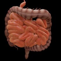
It is now known that, at the nervous system level, people with PE have an alteration in the substance nigra, a brain region that, among other functions, regulates movement control. In the context of this pathology, in this area there is a massive death of neurons that produce dopamine, a neurotransmitter associated with motor motivation and control. Unfishedyedly, in recent years various scientific research indicates that the gut would play a fundamental role in the death of these neurons. In this sense, alterations in the bacterial population that normally colonizes our intestine would be a favorable condition to induce the onset of the disease. Even if the bacterial population is relevant, a recent publication in the journal Nature adds a new edge to the proposal that brain-gut communication is key in PE.
Intestinal infections
Last June, Michel Desjardins’ laboratory in Canada showed that mice genetically predisposed to generating PE had the characteristic symptoms of the disease only when exposed to an intestinal infection. In particular, scientists took an interest in the study of the mitochondrial protein PINK1 which, by presenting a mutation in mice, unlike humans, generates mild symptoms of the disease. This led researchers to think that this genetic mutation alone was not enough to generate the pathology, but would require an additional factor.
Previous work by this same laboratory showed that infecting mice that did not express PINK1 with bacteria, which on their outer membrane have LPS (structure that allows some bacteria to withstand high temperatures, among other functions) induced that certain mitochondrial proteins were recognized by the immune system as a foreign agent, that is, a mechanism similar to that produced by autoimmune diseases. Because of this, the authors of this paper wondered whether a similar mechanism could kill the dopaminergical neurons of the substance nigra and generate THE EP.
Initially, researchers through food inoculated mutant mice for PINK1 with the bacteria Citrobacter rodentium, an intestinal pathogen that expresses LPS and induces a high autoimmune response against mitochondrial proteins. Once infected, the type of immune cells that recognized mitochondria in the spleen region was evaluated. To do this, they used flow cytometry, a technique that allows to count and classify cells according to their morphological characteristics and types of protein that they express on their surface. Through this methodology, it was observed that some of the immune system cells had on their surface, a receptor that responded to signals from a protein released by neurons. Thus, they suggested that if a neuron released such a protein it could attract immune system cells, which by detecting certain mitochondrial components on the surface of neurons could induce cell death.
To evaluate this hypothesis, they separated the mutant mice from PINK1 into two groups, one of which was inoculated with the bacteria C. rodentium and the other did not, and after that they performed a flow cytometry, but this time with the cells of the central nervous system. In this way, it was observed that only in the infected group, immune system cells traveled to the brain, recognized mitochondrial proteins on the membrane surface of neurons and consequently induced cell death. To itself, when they infected mice that normally expressed PINK1, immune system cells did not travel to the brain, so both the mitochondrial protein mutation and the infection were necessary to generate this recruitment. Then, to determine what type of neurons were affected, genetically modified mice whose immune system cells would only attack neurons that carry mitochondrial proteins on their surface. Interestingly, by isolating the neurons of these mice into a plaque and incubating them with LPS and immune system proteins, there was only one death of neurons that produce dopamine, that is, the cells responsible for generating EP.
Finally, the researchers assessed whether the intestinal infection induced motor disturbances in mice that did not express PINK1. To do this, they used the “pulley test” which consists of placing a mouse inverted position at the top of a mast 50 centimeters high and measuring the time it takes to rotate to a proper position and descend. This test is crucial for evaluating the symptoms of PE in mice, as it allows to measure motor control dependent on dopaminergic neurons. In this context, scientists demonstrated that mutant mice for PINK1, being inoculated with the gut bacteria, took longer to spin on the mast compared to the uned infected ones. Along with this, administering dopamine reversed the motor impairment in infected mice. Thus, in mice the absence of PINK1 is not enough to generate the symptoms of PE, but requires an intestinal infection through a bacterium that expresses LPS to generate Parkinson’s symptoms.
Brain-gut axis
This scientific finding, while not proposing a cure for PE, suggests for the first time that it could have an autoimmune origin. In addition, it provides a model to characterize the beginning of this pathology and eventually have certain therapeutic approaches. In this sense, decreasing the likelihood of intestinal infections and slowing down the immune response would be relevant to stop the progression of the disease early. However, this research was conducted on mice and therefore clinical trials are necessary to determine whether a similar mechanism generates pathology in humans.
Moreover, this work complements previous proposals that indicate that the normal functioning of the brain-gut axis is relevant to avoid some neurodegenerative diseases such as Alzheimer’s. In this way, it seems that the bacterial population that colonizes our intestine, as well as our defenses through the immune system are essential to regulate, in some way, the normal functioning of the nervous system. After all, not all the answers are in our heads…
Article Link: https://www.nature.com/articles/s41586-019-1405-y
*This article comes from the agreement with the Interdisciplinary Neuroscience Center of the University of Valparaiso


