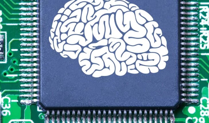
The development of new drugs is a complex and multi-stage process that involves a lot of resources but, above all, a large investment of time and money. Typically, after identifying potential therapeutic targets and developing candidate compounds, the latter are evaluated in cell cultures and animal models to determine their safety and efficacy before continuing with human clinical trials. But here’s the problem: both crops and animals have limitations related to their poor ability to mimic the conditions of the human body. On the one hand, the former cannot replicate all aspects of tissue function, that is, they do not behave as the liver, kidney or heart does within the body. This sometimes makes a drug that works well in the laboratory not effective when tested in people. With the latter, on the other hand, small genetic differences between species can be amplified to large physiological differences, since there are differences between the reaction of an animal and a human organism to the same stimulus. To address this problem, an alternative is “organs on chips”, devices the size of a pendrive made from human cells (from the heart, lungs, or brain, for example) that recreate on a very small scale the physical properties of an organ. The researchers believe that, with further development, they can help study diseases and test drugs in conditions closer to real life. The idea of organ-on-chips (OoCs) emerged in the 2000s, when the U.S. National Institutes of Health (NIH) in collaboration with the Food and Drug Administration (FDA) began funding the development of technologies to create accurate models of human organs in the lab. The NIH subsequently launched a program that aimed to develop 10 organs on different chips within 10 years. These consist of transparent channels lined with thousands of living cells and pumped with nutrient-containing fluid, or blood, all interacting just as they would in the body. Not only do they mimic blood flow, but they have microcameras that allow researchers to integrate multiple cell types to mimic the diversity of cells normally present in an organ. In turn, the presence of fluid makes it possible to connect these multiple cell types to study the interaction between them, as well as mimic what a cell experiences in the body, how it receives nutrients and removes waste, and how a drug will move in the blood. The ability to control fluid flow also makes it possible to adjust the optimal dosage for a particular medication. In 2014, the first successful case – which mimicked a lung – was presented and consisted of a layer of human lung cells grown on a silicone chip. This chip is capable of mimicking the dilation and contraction, or inhalation and exhalation, of the lung and simulating the interface between the lung and air. The ability to replicate these qualities made it possible to better study lung deterioration through different factors, and also the response of lung cells to different drugs and toxic substances. As technology advanced, other variations such as the liver, heart, intestine and kidneys were developed. On the other hand, current models of OoCs are difficult to use and have some disadvantages: creating them can be very complex and requires a high level of technical skill, require high investments of money, require living cells to function (and the availability of human cells may be limited in some cases), being living systems their operation can vary between different experiments, which may make it difficult to reproducibly the results; They are small systems and may not be scalable to a size suitable for human studies, and their regulation is under development, so it may vary between countries. In contrast, their advantages are also several: they can improve the safety and efficacy of new drugs before they are tested in humans, they can study human diseases under controlled conditions, they can be used to study how different patients respond to drugs (allowing the personalization of treatments, reducing the costs of drug research and development, Since they allow testing under controlled conditions before moving on to human studies, they serve to study biological processes and interactions between different body systems and can be used in theTo study rare diseases that are difficult to replicate in animal models. In short, organs on chips have great potential to improve the safety and efficacy of drugs, study human diseases, personalize treatments, reduce costs, and improve basic and toxicology research.
Organs on chips and their potential for the development of new drugs
January 18, 2023 |


